


This Article is part of the Computer Vision Resource Center.
A year ago, “medical imaging AI” was still mostly a slide-deck phrase — something vendors put in front of hospital boards next to a hockey-stick chart. In the meantime, however, this is no longer the case. For 2026, this is simply a line item. It has its own CPT codes. It has a regulatory deadline attached to a specific calendar date, not a vague “coming soon.” And it has, for the first time, a real evidence base showing where it actually moves the needle and where it’s still catching up to its own marketing.
Our broader guide on computer vision in healthcare covers the full landscape — surgery, ICU monitoring, pathology, the whole picture. This will get much more specific and focus wholly on imaging: the CT, MRI, X-ray, ultrasound and mammography AI that radiology departments are purchasing, billing and identifying with currently. Whether you are an administrator, developing a business case, a MedTech developer preparing your 2027 vision, or a radiologist seeking to assess the tool your hospital just bought.
Table of Contents
Key Takeaways
- Medical imaging AI has moved beyond pilot projects, with reimbursement pathways now available for select imaging applications.
- Radiology continues to dominate FDA-cleared AI medical devices, but only a small percentage have prospective randomized clinical evidence.
- Foundation models, vision-language models, and self-supervised learning are replacing many traditional single-task AI systems.
- The FDA’s Predetermined Change Control Plans (PCCPs) and the EU AI Act are reshaping how imaging AI products are developed and maintained.
- Hospitals should evaluate AI vendors based on clinical validation, interoperability, reimbursement potential, and long-term monitoring—not accuracy claims alone.
Let’s Get the Market Numbers Right First
Before anything else — because a lot of what’s floating around right now is wrong.
You’ve probably seen a headline claiming the medical imaging AI market is racing toward $20-plus trillion by 2035. It isn’t. That figure traces back to a single market research report that appears to have badly mislabeled its units (or conflated the AI segment with the entire global imaging hardware and services economy). Every other research firm covering the same category — Market Research Future, SNS Insider, Nova One Advisor, Roots Analysis — lands somewhere in the neighborhood of $18 billion to $38 billion globally by 2035, growing at a compound annual rate somewhere between 17% and 35% depending on how narrowly “AI in medical imaging” is defined. That‘s still a sizable growth story. Its just not a trillion dollar one and a hunk like this one loses credibility quickly if it is repeating a number that would not hold up for five minutes in a cross check.
What’s driving that growth isn’t mysterious. Imaging volumes keep climbing, radiologist shortages aren’t closing, and — this is the part that changed in 2026 — AI-enabled imaging analysis finally has real reimbursement pathways in some specialties, which is usually the thing that turns a pilot program into a purchase order.
The FDA Numbers, and the Gap Nobody Likes Talking About
As of the end of 2025, the FDA has cleared 1,451 AI-enabled medical devices, with 295 of those clearances happening in 2025 alone — the biggest single-year total on record. Radiology dominates that list. Roughly 1,104 of those clearances, about 76%, are imaging-related, dwarfing cardiology (9%) and neurology (5%).
Here’s the number that matters more, though: only about 2.4% of those cleared devices have been validated through a randomized controlled trial. The overwhelming majority — something like nine in ten — rely on retrospective data or have no published clinical evidence attached to them at all. That’s not necessarily a scandal. Most cleared devices go through the FDA’s 510(k) pathway, which asks a manufacturer to demonstrate “substantial equivalence” to an existing device, not to prove it improves outcomes in your hospital, with your patient population, on your scanners. A device may be legal on the market and yet perform very poorly once it is used in your case mix.
What does it mean if you are buying imaging AI in 2026? It means you have a commercially available device. Clearance simply means it has crossed a certain hurdle. It does not tell you the tool works well for you. Site-specific, prospective validation before go-live isn’t bureaucratic caution — it’s the only way to find out whether a model trained on someone else’s scanners and someone else’s patients holds up on yours.
The Regulatory Landscape Just Moved — Here’s What Actually Applies Now
This is a section worth reading carefully, because a lot of content published even a few months ago is already out of date on this.
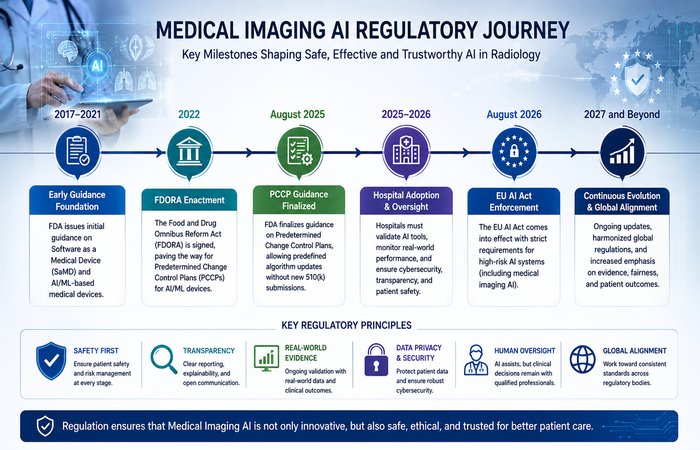
On the FDA side, the agency finalized its guidance on Predetermined Change Control Plans (PCCPs) in August 2025, giving statutory teeth to something Congress had authorized back in 2022 under the FDORA reform act. Here’s why this matters more for imaging AI than almost any other device category: unlike a physical implant, an imaging algorithm is built to keep learning. Historically, a meaningful update to a model — retraining it on new data, adapting it to a new scanner generation, refining it for an underrepresented patient subgroup — could trigger an entirely new 510(k) or PMA submission, sometimes taking months, during which patients were stuck using the older, worse version of the tool.
A PCCP changes that. Manufacturers now submit, upfront, a plan with three required components: a description of the specific modifications they intend to make, the protocol they’ll use to implement and test those changes, and an assessment of the impact on safety and effectiveness. If the FDA authorizes the plan, the manufacturer can roll out those pre-specified updates without going back for a new submission each time. It’s a real shift toward treating AI/ML software the way it actually behaves — as something iterative — rather than forcing it into a regulatory framework built for a device that doesn’t change once it ships. Alongside this, the FDA’s Quality Management System Regulation (QMSR), which aligns US device quality requirements with ISO 13485, becomes effective February 2, 2026, adding another layer manufacturers need to fold into their existing processes.
On the EU side, the timeline has genuinely changed, and this is where a lot of 2026 content is going to age badly if it isn’t corrected. The original AI Act delayed high-risk requirements to 2 August 2026 for stand-alone AI and to 2 August 2027 for integrated ones (in regulated products like medical devices); implementation was apparently falling at least a year further behind schedule for most of 2025, which prompted the European Commission to resurrect the ‘Digital Omnibus on AI’ in November 2025 to shelve the operative dates. After a turbulent first trilogue in April 2026 that fell through for lack of consensus, the negotiators achieved a political agreement on 7 May and the EU Council ratified it on 29 June 2026.
The confirmed, currently binding dates are:
- December 2, 2027 — high-risk obligations apply to stand-alone Annex III systems (the use-case-driven category covering things like biometric or employment AI).
- August 2, 2028 — high-risk obligations apply to AI embedded in products already regulated under existing EU product-safety law, which is the category medical imaging AI falls into as a component of MDR- or IVDR-regulated devices.
- December 2, 2026 — the Article 50 transparency and AI-generated-content watermarking obligations, which weren’t part of the delay and stayed roughly on schedule (with a short grace period added for legacy systems).
If this is a compliance roadmap you’re working on, you should be planning to target August 2, 2028, for medical imaging AI, not the August 2027 date (which is still floating around in a lot of places). And don’t read the extension as a pass — every law firm and regulatory consultancy tracking this agrees on one point: the extra runway exists because the standards bodies and Notified Bodies weren’t ready, not because the underlying obligations got lighter. Those manufacturers who begin their risk classification, technical documentation and data governance work now will progress through certification queues that are orders of magnitude faster than those who wait until 2027.
One structural aspect good to keep in mind when selling into both the US and EU: Article 8 of the AI Act enables one Notified Body to undertake both the conformity with the MDR/IVDR and the high-risk requirements of the AI Act in one assessment process, rather than a dual one. Paired with the fact that Class I devices not requiring Notified Body review under MDR generally fall outside the AI Act’s high-risk bucket unless specifically listed under Annex III, this gives compliance teams a real lever to prioritize budget toward the diagnostic and therapeutic product lines that actually carry the regulatory weight.
From Narrow Models to Generalists: The Architecture Shift
For most of the last decade, medical imaging AI meant one model trained for one job — a network that found lung nodules and did nothing else, built on thousands of hand-labeled images that took a small army of radiologists to annotate. That’s changing, and it’s changing for a specific technical reason: labeled medical data is expensive and slow to produce, and it’s always been the bottleneck.
Two things are chipping away at that bottleneck. The first is the rise of foundation models purpose-built for medical images. Meta’s Segment Anything Model (SAM) and its medical adaptation, MedSAM — trained on more than 1.5 million image-mask pairs — represent a genuine architectural departure from the single-task model. MedSAM’s 0.914 median Dice score on vertebrae segmentation is competitive with specialist models trained for that job alone, which is the whole point: a generalist model that can be adapted to many tasks with comparatively little additional training data. Vision Transformer architectures are following a similar trajectory — models like VerFormer are already outperforming conventional CNNs on multicenter spine benchmarks by building in attention mechanisms tuned to anatomical structure rather than generic image patches.
The second shift is self-supervised learning, which sidesteps the labeling bottleneck almost entirely by letting a model learn useful representations from unlabeled images before it’s fine-tuned on a much smaller labeled set for a specific clinical task. The emerging refinement — sometimes called task-aligned SSL — tries to get the best of both: the scale of unsupervised pretraining with the precision of supervised fine-tuning.
Then there’s the frontier that’s arguably the most consequential for how radiologists actually work day to day: vision-language models. These don’t just flag an abnormality — they generate structured clinical language describing what they found, integrating the visual finding with context from the patient’s history. The first foundation-model-powered devices built on this approach are already reaching FDA clearance, and the practical implication is faster deployment: a model built this way can, in principle, adapt to a new hospital’s imaging protocols in weeks through zero-shot or few-shot adjustment, rather than requiring years of supervised retraining the way older single-task models did.
None of this replaces the radiologist’s report. What it changes is the starting point — a drafted, structured description the radiologist edits and confirms, rather than a blank worklist.
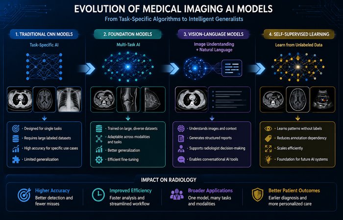
Comparison of Major Medical Imaging AI Approaches
| Approach | Primary Use | Strengths | Limitations |
| Traditional CNN Models | Single disease detection | High accuracy for specific tasks | Requires extensive labeled datasets |
| Foundation Models | Multiple imaging tasks | Adaptable across specialties with minimal retraining | Still undergoing broader clinical validation |
| Vision-Language Models | Image interpretation and report generation | Creates structured clinical reports | Requires strong governance and human oversight |
| Self-Supervised Learning | Training using unlabeled images | Reduces dependence on manual annotation | Fine-tuning is still required for specific clinical applications |
The Reimbursement Story Nobody Was Talking About a Year Ago
If there’s one development that separates 2026 from prior years, it’s this: medical imaging AI finally has a real, working reimbursement pathway in at least one specialty, and it’s changing purchasing behavior in a way that survey data and pilot programs never did.
On 1 January 2026, CPT code 75577 became a permanent Category I code for AI-based quantification and characterization of coronary plaque from coronary CT angiography (CCTA) succeeding the temporary Category III codes (0623T–0626T) under which vendor were billing for years. The reimbursement figures are (not surprisingly) more substantial: CMS assigned an Hospital Outpatient Prospective Payment System (OPPS) rate of $950.50 and a Physician Fee Schedule rate of a little over $1,000 per case (Elucid reports a national average of $1,012–$1,021 depending upon setting). Major commercial payers — Aetna, UnitedHealthcare, Cigna, and Humana among them — moved to cover it around the same time, extending access to tens of millions of commercially insured patients on top of Medicare coverage.
Companies like Cleerly (first FDA clearance in this category, back in 2020), HeartFlow, Elucid, Caristo Diagnostics, Artrya, and Circle Cardiovascular Imaging have all built products around this. What’s instructive about how they got here is the point made repeatedly by clinicians involved in the CPT process: the codes exist because these companies invested heavily in clinical outcome studies, not just accuracy benchmarks. As Eric Rubin, MD — the American College of Radiology’s CPT advisor to the AMA — put it in discussions at RSNA 2025, plaque analysis and FFR-CT earned Category I status specifically because their sponsors anchored the technology to evidence of patient impact, not just technical performance metrics like AUC. That’s a template other imaging AI categories will likely need to follow if they want their own permanent codes rather than perpetually renewed temporary ones.
Outside cardiac CT, reimbursement for most AI-enabled imaging services remains murky. There’s still no established CPT pathway for the majority of AI-assisted radiology functions — triage prioritization, most segmentation and detection tools, and general diagnostic support software largely operate without a dedicated billing code, which means the business case for them still has to be built on cost avoidance and workflow efficiency rather than direct revenue. One frequently cited health-economic model puts the incremental cost-effectiveness of AI-assisted vertebral fracture screening at roughly $72,000 per quality-adjusted life year — a number worth having in your back pocket if you’re building an ROI case, because it’s one of the few concrete figures available rather than a vendor projection.
Leading Medical Imaging AI Vendors in 2026
| Company | Primary Focus | Notable Technology |
| HeartFlow | Cardiac CT Analysis | FFR-CT and coronary plaque assessment |
| Cleerly | Coronary artery disease | AI-powered plaque characterization |
| Aidoc | Radiology workflow | Critical findings detection and prioritization |
| Zebra Medical Vision | Multi-condition imaging AI | Chest, liver, bone and cardiovascular analysis |
| Subtle Medical | MRI and PET enhancement | Image quality improvement with faster scans |
The Evidence Base Is Finally Catching Up — A Few Studies Worth Knowing
Skepticism about imaging AI’s evidence base is warranted, but it would be inaccurate to say nothing has changed. A handful of 2025–2026 studies are genuinely worth knowing by name, because they represent the kind of peer-reviewed, outcomes-anchored evidence the field has been missing.
The clearest example is UltraSight’s cardiac ultrasound work. In a multicenter trial published in the European Heart Journal – Digital Health, 2026 (DOI: 10.1093/ehjdh/ztag065), nine new users a mix of 6 nurses and 3 medical students with no pre-existing knowledge of ultrasound were trained in a video-led simulation session for about 8 hours on AI-assisted image acquisition before being left on their own to scan real patients. Of 954 novice-acquired images, 97.7% met the diagnostic-quality threshold, and every operator hit diagnostic-quality scores from their very first independent scan. A related companion study at the same journal, testing the same AI-guidance approach across 496 patients, found expert reviewers could reliably rule out left ventricular dysfunction in 99.4% of cases and left ventricular hypertrophy in 98.7% of cases using the novice-acquired, AI-guided images. That’s a meaningful result for any health system trying to expand echocardiography access beyond centers that can staff a trained sonographer.
The point of citing this isn’t that AI-guided ultrasound is a silver bullet — it’s that this is what real clinical evidence looks like: named authors, a registered trial, a specific effect size, and a journal DOI you can go check yourself. That’s the standard the rest of the imaging AI market still needs to catch up to, and it’s worth demanding it from any vendor pitching you a similar claim without the paper to back it up.
Interoperability: Where Most Deployments Actually Die
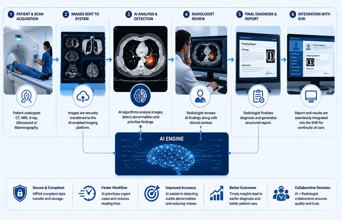
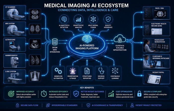
Ask anyone who’s run a failed imaging AI rollout what went wrong, and the answer is rarely “the model wasn’t accurate enough.” It’s almost always integration. A model can hit impressive benchmark numbers in a vendor’s lab and still be useless in production if it can’t talk to the systems radiologists already live in — PACS, RIS, and the EHR.
The practical fix is building on open standards from day one rather than retrofitting them later. DICOMweb has become the modern standard for programmatic access to imaging data over standard web protocols, replacing older point-to-point DICOM integrations that required custom interfaces for every new system pairing. On the clinical-data side, FHIR’s ImagingStudy resource gives a standardized way to represent metadata about an imaging exam — modality, body site, series, and study identifiers — in a format that can be queried and exchanged the same way any other clinical resource is, closing the gap between a pixel-level finding and the patient’s broader chart. Get this wrong, and even a highly accurate model becomes a second, disconnected interface that adds clicks instead of removing them — which is exactly the kind of friction that accelerates the clinician burnout the tool was supposed to reduce in the first place.
The Shift Toward Agentic Workflows
The other genuinely new theme in 2026 is the move from AI that waits for a prompt to AI that acts with a goal. In radiology specifically, “agentic” systems are starting to show up doing three things without a human initiating each step: organizing a case before a radiologist opens it (pulling relevant prior studies and flagging what’s changed), surfacing missing patient history or lab results that are sitting in a disconnected EHR silo, and automating the follow-up coordination after a finding is confirmed — routing a critical result to the right care team without someone manually picking up the phone.
This isn’t decision support in the traditional sense — it’s closer to a proactive coordinator sitting alongside the radiologist. It also creates a governance question hospitals haven’t fully solved yet: agentic systems still need a competent human supervisor in the loop, and that’s beginning to show up as a distinct job function — an AI quality assurance role responsible for monitoring what these systems are doing autonomously, not just what they recommend.
This capability compounds when paired with longitudinal patient data — a continuous record that follows a patient from the ICU to a general ward to virtual care and eventually home, rather than a series of disconnected imaging snapshots. An agentic system that can see a patient’s imaging trend over time, not just today’s scan, is a meaningfully different tool than one reading a single study in isolation. Getting there depends on the same open-standards foundation described above; longitudinal value dies quickly in a proprietary, closed data silo.
What Medical Imaging AI Will Look Like Beyond 2026
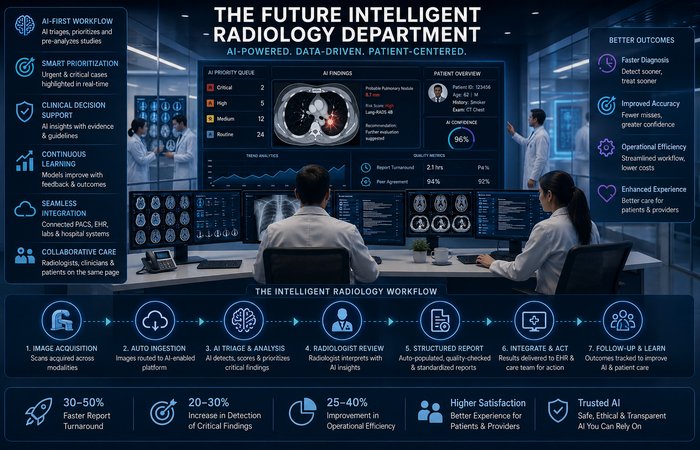
Over the next several years, medical imaging AI is expected to evolve from isolated diagnostic tools into integrated clinical assistants. Instead of simply identifying abnormalities, future systems will combine imaging findings with laboratory results, electronic health records, genomic data, and longitudinal patient histories to support more personalized clinical decisions.
Hospitals are also expected to adopt increasingly autonomous workflow assistants that prioritize cases, coordinate follow-up care, monitor quality metrics, and continuously evaluate model performance. As foundation models mature and regulatory frameworks become more standardized, medical imaging AI will likely become part of routine clinical infrastructure rather than an optional technology investment.
A Practical Checklist Before You Sign Anything
Pulling this together into something usable — if you’re evaluating a medical imaging AI vendor in 2026, these are the questions worth insisting on answers to before you commit budget:
- What’s the actual evidence? Ask specifically whether performance claims come from a prospective study or a retrospective one, and whether it’s been peer-reviewed with a named journal and DOI — not a white paper.
- Does it hold up on your patient population and your scanners? Aggregate accuracy numbers can hide serious subgroup disparities across age, sex, ethnicity, and equipment vendor. Insist on subgroup breakdowns, and if the vendor can’t provide them, treat that as a serious flag rather than an oversight.
- Does it have a PCCP, and what’s actually in it? For FDA-cleared devices, ask whether the manufacturer has an authorized Predetermined Change Control Plan and what specific modifications it covers — this tells you how the tool is going to evolve after you buy it, and under what oversight.
- What’s the reimbursement pathway, if any? Outside of cardiac CT plaque analysis and FFR-CT, most imaging AI still doesn’t have a dedicated CPT code. Know upfront whether you’re building the business case on billing revenue or on workflow savings, because those require very different financial models.
- How does it integrate — really? Confirm DICOMweb and FHIR support explicitly, and ask to see it working inside your actual PACS/RIS environment before go-live, not in a vendor sandbox.
- Who owns drift monitoring after deployment? Get a documented answer on how performance is tracked over time and what triggers retraining or re-evaluation as your patient mix or equipment changes.
- What’s your EU exposure, and are you planning against August 2028? If you sell or deploy in the EU, confirm the vendor’s compliance roadmap is built around the confirmed Digital Omnibus dates, not the original 2027 timeline.
FAQs
Q1: Can AI replace radiologists?
A: No. Current medical imaging AI is designed to assist radiologists by identifying abnormalities, prioritizing urgent cases, and generating preliminary findings. Final diagnosis and clinical decision-making remain the responsibility of qualified healthcare professionals.
Q2: Which imaging modalities benefit most from AI?
A: Computed tomography (CT), magnetic resonance imaging (MRI), chest X-rays, mammography, and cardiac imaging currently have the most mature AI applications and the largest number of FDA-cleared solutions.
Q3: What is a Predetermined Change Control Plan (PCCP)?
A: A PCCP allows AI device manufacturers to make pre-approved algorithm updates under an FDA-authorized plan without submitting a completely new regulatory application for every modification.
Q4: Is every FDA-cleared AI system clinically proven?
A: No. FDA clearance confirms that a device meets regulatory requirements, but hospitals should also evaluate prospective clinical validation, peer-reviewed evidence, and real-world performance before deployment.
Q5: What should hospitals consider before purchasing imaging AI?
A: Organizations should evaluate clinical evidence, interoperability with PACS and EHR systems, reimbursement opportunities, workflow integration, cybersecurity, regulatory compliance, and long-term performance monitoring.
Where This Actually Leaves Us
The honest version of the 2026 story is less dramatic than the market-size headlines suggest, and more interesting because of it. Medical imaging AI didn’t suddenly become trillion-dollar technology this year. What actually happened is quieter and more durable: a real reimbursement code went live and proved the economics can work when the evidence is strong enough to justify it; a regulatory framework on both sides of the Atlantic got more concrete, not less, even as the deadlines moved; and the architecture underneath the tools started shifting from thousands of narrow, single-purpose models toward a smaller number of adaptable, multimodal ones.
None of that closes the evidence gap on its own. Most cleared devices still haven’t been through a randomized trial, and most AI-assisted imaging work still doesn’t have a billing code attached to it. But the direction is toward more rigor, not less — and for an industry that spent the better part of a decade being judged mostly on demo accuracy, that’s the more important trend to be watching in 2027.
Related Guides