
This Article is a part of
Computer Vision Resource Center
Walk into any busy radiology department today and you will find a silent crisis in progress. Radiologists are reviewing 50 to 100 scans per shift. Surgeons are doing more complex procedures than ever before, on an aging patient population, with less support staff. And nurses? They spend just 37% of their shifts on actual, direct patient care.
Something has to give. And increasingly, that something is computer vision.
This isn’t hype anymore. Today the FDA has approved 1,451 medical devices with some form of AI; the pace of approval last year reached 295 devices, the most in any year up to 2025. Medical technology which a decade ago would only have been found in research labs is now becoming commonplace in operating rooms, intensive care units and imaging suites globally.
Here‘s the truth most people won‘t tell you: not all of those solutions are equally good, and using them the wrong way can lead to more harm than good. In this article, I‘ll highlight what computer vision can actually do in healthcare, where it has the potential to add real value, and the tough questions a hospital or MedTech developer needs to ask before using it.
Table of Contents
What Is Computer Vision in Healthcare?
Let‘s get on the very basics to start with as the terminology can become very muddled very quickly.
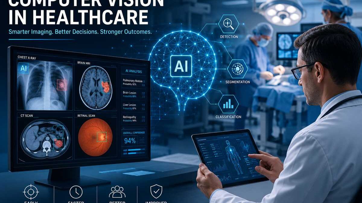
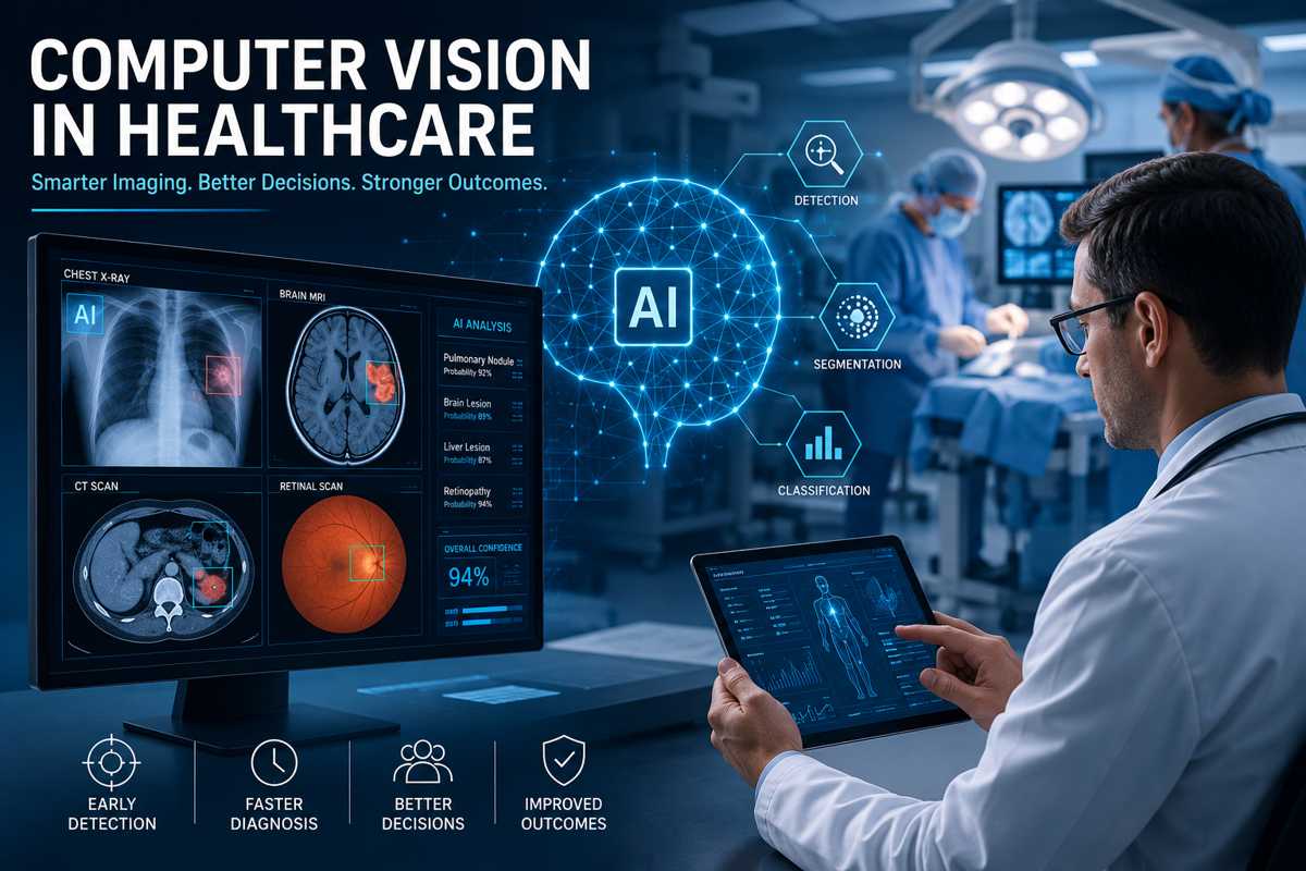
Computer vision (CV) is the area of AI that enables computers to understand the visual world X-rays, MRIs, CT scans, pathology slides, endoscopic videos, live camera feeds from hospital wards.
Computer vision in healthcare is one of the fastest-growing applications of visual AI technology. To understand the broader concepts, algorithms, and industries using this technology, explore our complete Computer Vision guide covering how machines interpret and analyze visual information.
Imagine it as clothing software in a set of expert clinical eyes.
To understand where it fits, it helps to see the full hierarchy:
- Artificial Intelligence (AI) — the broad field of machines performing tasks that normally require human thinking.
- Machine Learning (ML) — a subset of AI where algorithms learn from data instead of being explicitly programmed.
- Deep Learning (DL) — a form of ML that uses multi-layered neural networks to pull complex patterns out of messy, high-dimensional data.
- Computer Vision (CV) — the application of AI and deep learning specifically to visual
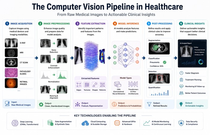
Most CV workflows in medicine share a recognizable pipeline:
- Image acquisition and preprocessing — Input into the registration algorithm through using one standard format, regardless of the specific scanner, hospital or imaging
- Core CV tasks — Segmentation (defines anatomy), detection (localizes the abnormality), classification( Categorize the anomaly into an appropriate diagnosis)and land marking (Measurement of the anatomy/abnormality and their relationship to each other).
- Clinical interpretation layer — converting raw model outputs into clinically meaningful measurements or flags.
- Workflow integration — Reporting of findings back into the current clinician systems (PACS, RIS, EHRs) with the necessary human review.
- Monitoring and governance — Must ensure that the performance doesn‘t degrade as equipment, patient groups, or clinical processes are modified over time.
The real magic isn’t just automation. It’s what specialists call high-order feature extraction — the ability to spot patterns in a pixel that no human eye could catch. A convolutional neural network (CNN) can detect the faintest hairline fracture, the earliest signs of a tumor, or a subtle change in a retinal scan that signals disease years before symptoms appear.
Accuracy alone, though, isn’t enough for clinical adoption. Repeatability, generalizability, latency, interoperability, and usability all matter — and all of them operate under regulatory and privacy constraints that get tighter every year.
That’s the promise and the challenge. Now let’s look at where it’s actually working.
How Does Computer Vision Work in Healthcare?
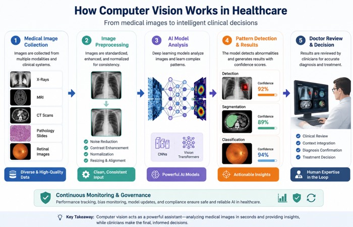
Computer vision in healthcare doesn’t think the way a clinician thinks. It doesn’t “see” a lung or a fractured vertebra the way a radiologist does. What it does instead is recognize patterns — millions of them — learned from enormous volumes of previously analyzed medical images. Understanding the pipeline behind that process helps clarify both its power and its limitations.
Modern computer vision models are becoming more advanced by combining image recognition with language understanding. Similar techniques are used in automatic image and video caption generation with deep learning, where AI systems analyze visual content and generate meaningful descriptions.
Medical Image Collection
Everything starts with data. Other data may include CT scans, MRIs, X-rays, pathology slides, endoscopic feeds, retinal photographs and so on, collected from clinical systems. The more varied the data the better the performance of the model will be in the long run. Training solely on images from one hospital, one scanner brand or even the same team of clinicians, the model must work through any number of seemingly random outliers.
Image Preprocessing
Raw medical images are rarely clean enough for direct analysis. Preprocessing standardizes brightness, contrast, resolution, and orientation across images captured on different equipment under different protocols. This step removes noise, corrects artifacts, and ensures the model receives consistent input — because inconsistent input produces unreliable output.
AI Model Analysis
The input image has been pre-processed and been entered into a deep learning model, such as a CNN or increasingly, a Vision Transformer model. The input image is dissected into pixel-level patterns for the model to analyse in relation to all training data it has seen.
An AI reading a chest x-ray does not have the same perceptual experience as a human physician. It is “perceiving” arrays of learned pixel pattern “features” statistically correlated with pneumothorax, fractures, or tumors.
Pattern Detection
Confidence measures used to highlight suspicious areas that pattern is linked to. i.e. abnormality in segmentation, classification of normal/abnormal, locating abnormality. The outcome of a pattern match can be segmentation map, bounding box of abnormality, classification label.
Doctor Review
No cleared clinical CV system operates without a human in the loop. The model’s output reaches the clinician — through PACS, RIS, or EHR — as a decision-support signal, not a final diagnosis. The physician reviews the flagged findings, applies clinical judgment, and makes the call. The AI narrows the search. The doctor owns the conclusion.
Solving the Imaging Crisis: High-Throughput Diagnostics and Early Detection
Radiology is where computer vision has made its biggest dent, and for good reason. It accounts for 76% of all FDA-cleared AI devices — 1,104 in total — far ahead of cardiology at 9% and neurology at 5%.
The core problem CV solves here is volume. When a radiologist is staring down a queue of 100 scans, the danger isn’t lack of skill — it’s fatigue and backlog. A critical finding buried in scan number 87 might not get flagged for hours. In time critical situations such as intracranial hemorrhage or tension pneumothorax, those hours count significantly.
AI as a Force Multiplier
Rather than replacing radiologists, well-designed CV systems act as triage assistants. They scan the entire queue in seconds and push the urgent cases to the top for immediate review. The radiologist still makes the call. The AI only writes that the most ill patient isn‘t kept waiting for the more semi-urgent studies.
Even minor prioritization enhancements produce downstream clinical benefit: quicker escalation to ED nursing/MD, earlier time-to-therapy, and fewer silent delays instigated by the backlog.
The operational win is strongest when CV outputs appear directly in PACS and RIS — where clinicians already work — rather than in a separate disconnected interface.
Where the Accuracy Numbers Actually Land
The performance data across imaging tasks is genuinely impressive:
| Application | Model | Performance |
| Osteoporotic fracture grading | YOLO-XRAY | 96.9% accuracy (external validation) |
| Vertebral segmentation | MedSAM | 0.914 median Dice score |
| Disc narrowing and stenosis | SpineNet | Matches human radiological grading |
| Tumor differentiation (schwannoma vs. meningioma) | CV model | AUC 0.876 |
| Intramedullary tumor vs. demyelinating lesion | CV model | 96% accuracy |
| Hardware failure detection (rod fractures) | CV-assisted radiologist | Recall rate: 0.70 → 0.95 |
Beyond Bones: Ophthalmology and Digital Pathology
Radiology grabs the headlines, but two other fields deserve attention because they represent some of computer vision’s cleanest clinical wins.
In ophthalmology, CV systems are now used to perform automated screening for diabetic retinopathy, one of the world‘s main causes of preventable blindness. Such screening makes possible early detection in primary-care clinics, without waiting for a specialist referral that could be many months distant. Timely triage here doesn’t just improve outcomes; it prevents irreversible progression.
In digital pathology, CV algorithms analyze tissue slides at a scale no human pathologist could match — flagging suspicious regions on biopsy samples, supporting tumor localization, and helping standardize a discipline that has long suffered from inter-observer variability. The challenge isn’t only model accuracy. Variability between slide scanners, staining procedures, and lab workflows impact performance in the real world.
What unites radiology, ophthalmology, and pathology? Consistency. A tired human varies. A validated algorithm doesn’t.
Inside the Intelligent OR: Real-Time AI Support for Surgery
If radiology is CV’s proving ground, the operating room is its toughest test — and arguably its highest-stakes payoff.
The OR is a brutal environment for any vision system. There’s bleeding. There’s surgical smoke. Instruments and hardware block the view. Anatomy shifts as the surgeon works. Getting reliable, real-time interpretation out of that chaos is a genuine engineering achievement — and it’s happening.
Real-World Examples of Computer Vision in Healthcare
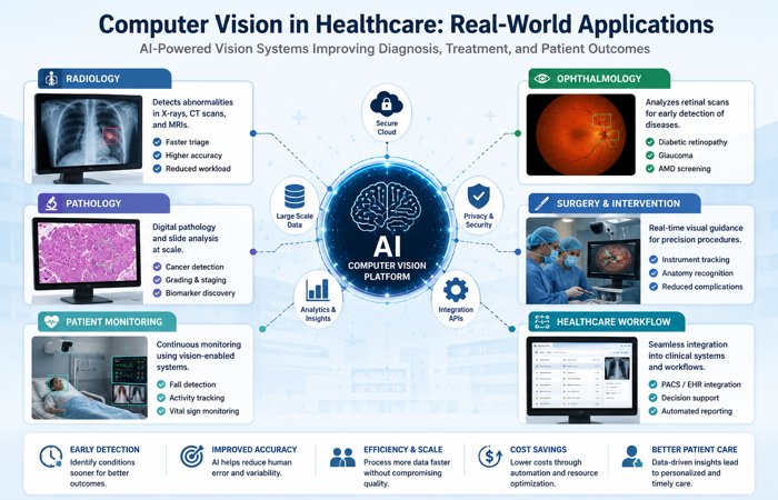
Understanding how computer vision performs in controlled research is one thing. Seeing how it operates inside real clinical environments — at scale, on real patients — is another. These five platforms represent the clearest proof points that computer vision in healthcare has moved well beyond the research lab.
Google AI — Diabetic Retinopathy Screening
DeepMind AI system developed by Google‘s DeepMind can identify detecting diabetic retinopathy and diabetic macular edema- two of the most common consequences for preventable blindness by analyzing retinal fundus images. The system was trained on more than 128,000 retinal images. During clinical validation, its sensitivity was comparable to that of board-certified ophthalmologists. In practice, this means a primary care clinic in a rural or underserved area — where a specialist may be months away — can run a retinal scan and receive an immediate, reliable screening result. Google’s system has been piloted across healthcare settings in India and Thailand, demonstrating that CV can close geographic gaps in specialist access that no amount of physician recruitment could realistically solve.
Aidoc — Radiology AI Triage
Aidoc is a backend intelligence layer within radiology departments that continuously screens diagnostic images as they arrive and surfaces time-sensitive findings at the top of the radiologists ’ worklist. The Aidoc platform analyzes images for pulomnary embolism, intracranial bleed, cervical spine fracture, and aortic dissection, among others. The platform does not supplant the radiologist‘s read; rather, it reprioritizes the worklist, bringing time-sensitive studies to the top of the radiologists ’ queue before he or she has even opened it. Aidoc has FDA clearance across multiple indications and is in use at hundreds of hospitals worldwide. Published experience with Aidoc has demonstrated a decline in time-to-diagnosis for time-sensitive diagnosis’ – translating into patient survival and reduced long-term disability in conditions such as stroke and pulmonary embolism.
While computer vision focuses on interpreting visual medical data, AI diagnostics expands those capabilities by combining imaging with laboratory results, patient histories, and intelligent clinical workflows. Read our AI Diagnostics guide to see the bigger picture.
Viz.ai — Stroke Detection
Each minute delay in stroke treatment, cost the patient 1.9 million neurons. Viz.ai was designed with that in mind. The platform employs computer vision algorithms to analyze a patient’s CT angiography, in real time, and autonomously determine if a large vessel occlusion (LVO) the most severe but time-dependent stroke is present. An mobile alert instantly activates the appropriate specialist as opposed to waiting on a radiologist interpretation, flag to the neurology team, and manual call. This entire time-consuming sequence of events is condensed into a matter of minutes. The platform is FDA De Novo approved and peer-reviewed literature demonstrates the time from imaging to specialist notification dropped by greater than 50%. The product is currently implemented in some of the largest stroke systems in the US and Europe.
PathAI — Digital Pathology
Pathology has traditionally been one of medicine‘s most subjective fields. Two experienced pathologists looking at a slide may disagree. In oncology, the stakes for consensus are high disagreements can mean the difference between Type I and Type II chemotherapy. PathAI uses computer vision on large databases of digitized pathology slides to help pathologists with AI-assisted tissue morphology classification, tumor region detection, biomarker quantification and disease grading. PathAI‘s models are not designed to replace the pathologist‘s decision they are a second set of eyes to provide more evidence, trained on larger datasets than one practitioner can process in their lifetime. The company has partnered with major pharmaceutical organizations and academic medical centers to deploy its platform across oncology workflows, where consistent, reproducible slide interpretation directly affects clinical trial quality and treatment precision.
NVIDIA Clara — Medical Imaging AI Platform
Clara is not just one clinical application rather, it is the infrastructure tier upon which several applications run. It is NVIDIA‘s comprehensive platform for medical imaging AI, offering the compute framework, pre-trained models, federated learning tools and deployment tools that hospitals and MedTech companies utilize to develop and deploy clinical CV tools. Its federated learning feature is noteworthy: it enables hospitals to team up and train AI models without sharing patient data externally, working around the longstanding obstacles to multi-site AI development. Clara is compatible with CT, MRI, ultrasound and pathology imaging, and works with NVIDIA‘s GPU hardware to provide the high processing speeds necessary for real-time intraoperative and diagnostic settings.
What all of the above five examples have in common, and what all the students who wrote these examples have in common. None of them replace the clinician. Each one accelerates, standardizes, or extends what a skilled human team can accomplish — whether that means screening more patients, catching more critical findings faster, or bringing specialist-level interpretation to settings where specialists aren’t available. That is the real-world value proposition of computer vision in healthcare, and these platforms are delivering it today.
Many healthcare organizations now combine computer vision with advanced imaging AI platforms to improve diagnostic accuracy and streamline radiology workflows. Learn more in our Medical Imaging AI guide for radiology.
From Retrospective Review to Live Guidance
For years, surgical AI was limited to reviewing video after the fact — useful for training and skill assessment, but irrelevant to the moment that mattered. That’s changing. Modern intraoperative decision support systems now offer:
- Phase recognition — automatically identifying which stage of a procedure is underway, such as gallbladder retraction during a laparoscopic cholecystectomy. This enables automated skill assessment and triggers context-specific safety alerts.
- Anatomy identification — flagging critical structures like the ureter or common bile duct in real time so the surgeon doesn’t accidentally injure them.
- Instrument tracking — following tools relative to anatomy to support safety-zone alerts and reduce the margin for catastrophic error.
Endoscopic scene-understanding algorithms like Frame-UNet — achieving a 0.89 Dice score — give surgeons real-time visual awareness of delicate structures including nerves and the dura mater, functioning as an intraoperative safety net against neurological injury.
Augmented Reality and Marker-less Navigation
AR platforms like xVision project navigational guidance directly into the surgeon’s line of sight, achieving 94.1% accuracy in screw placement. No looking away at a separate monitor. The guidance appears right on the surgical field, exactly where attention already lives.
Traditionally, surgical navigation required ionizing radiation and physical tracking markers. Computer vision is quietly dismantling both requirements:
- Marker-less registration using U-Net architectures generates anatomical poses straight from preoperative images, achieving registration errors of just 1 mm — without a single physical marker placed in the patient.
- BoneMRI uses patch-based CNNs to synthesize CT-quality data from MRI scans, achieving a 0.84 Dice score. The result is a radiation-free navigation workflow that reduces exposure for both patients and surgical staff while maintaining submillimetric accuracy.
The Spine Surgery Case Study: What Mature, Commercial CV Looks Like
Spine surgery deserves its own spotlight because it illustrates what happens when CV moves beyond research into full commercial deployment.
Nearly one million adults in the United States undergo instrumented spinal procedures every year. Elective lumbar fusions increased by 62.3% between 2004 and 2015 — and by a staggering 138.7% among patients aged 65 and older. That surge in complex, high-cost procedures demands tools that can scale precision without scaling errors.
Commercial players have responded decisively. Medtronic (UNiD), ATEC (EOS Insight), and Carlsmed (Aprevo) now use CV-driven segmentation to create patient-specific rods and interbody cages. The outcome data is concrete: AI-driven custom implants have reduced rod fracture rates from a historical 9% down to just 2.2%.
On alignment measurement, CV now matches or beats human experts. IB Lab SQUIRREL, trained on over 17,000 images, agrees with specialist radiologists to within 0.16 degrees. SpinePose predicts landmarks with median errors as low as 1.3 degrees for pelvic tilt and 2.2 mm for Sagittal Vertical Axis. Multitask learning models are hitting intraclass correlation coefficients of 0.96 to 0.99 even in severe deformity cases — effectively outperforming many orthopedic surgeons on raw measurement accuracy.
Pervasive Patient Monitoring: Beyond the Manual Check
Here’s a use case most people never think about, but it may carry some of the highest real-world impact.
Hospital wards and ICUs still rely heavily on periodic manual checks. A nurse looks in, notes what she sees, moves on. Between those checks — which may be 30, 60, or 90 minutes apart — a lot can happen. A patient reaches for a rail and falls. The early, flickering signs of delirium appear and pass unnoticed. A change in posture signals deterioration that won’t be caught until the next round.
Computer vision offers something manual rounds simply cannot: continuous, passive surveillance.
Catching Falls and Delirium Early
Passive video-analysis platforms now monitor for the things humans miss between rounds. By tracking posture and motion continuously, these systems flag fall risk before a patient hits the floor. For ICU delirium — a condition with fluctuating symptoms that periodic checks routinely miss — CV analysis of posture and head-pose variation can identify at-risk patients with 94% accuracy.
Given that nurses spend only 37% of their shifts on direct patient care, always-on monitoring functions as a genuine staffing multiplier. It frees clinical teams to focus on the work only humans can do, while the system handles the vigilance that machines are better suited for.
The Privacy Question — and a Surprising Answer
The obvious concern: isn’t 24/7 video surveillance a HIPAA problem?
This is where the data becomes genuinely reassuring. The assumption has always been that strong privacy protections gut accuracy. But advanced monitoring platforms have proven otherwise. LookDeep Health’s Model v5 reached an F1-score of 0.91 even with patient faces fully blurred.
Think about what that means. You can obscure the single most identifying feature in the frame — the face — and the model still performs at a clinical-grade level for fall prevention and delirium detection. Privacy and performance are no longer a forced trade-off. With the right architecture, you get both.
The practical implication for hospital procurement teams: don’t accept the framing that HIPAA compliance requires sacrificing AI efficacy. Demand evidence of performance after privacy transforms have been applied — not before.
The Regulatory Hurdle and the Validation Gap
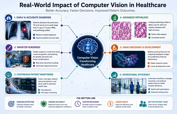
Now for the section that separates careful buyers from expensive mistakes.
The 1,451-device headline is exciting. But before you sign a purchase order, you need to understand what that number does not tell you.
The Uncomfortable Truth About “FDA-Cleared”
Most FDA-cleared AI devices go through the 510(k) pathway, which requires demonstrating “substantial equivalence” to a predicate device — not proof that the tool actually improves patient outcomes in your population. Most are classified as Software as a Medical Device (SaMD), subject to either 510(k) or De Novo classification depending on risk level and novelty.
The gap between “cleared” and “clinically proven” is wide and consequential:
- Roughly 90% of FDA-cleared AI devices rely on retrospective data or have no public clinical evidence at all.
- Only 2.4% have been validated through randomized controlled trials.
Read that again. Out of nearly 1,500 cleared devices, barely one in forty has been tested using the gold standard of clinical evidence.
This is the validation gap — and it is the single most important concept in this entire article for any organization considering purchasing or deploying medical CV. Market momentum is not clinical utility. A device can be cleared, marketed, and actively sold while still underperforming on your patient population, your imaging hardware, and your clinical workflow.
Why Site-Specific Validation Is Non-Negotiable
The single most protective thing any healthcare organization can do is run site-specific prospective validation before live deployment. Test the tool against your local patient demographics and your actual scanning equipment. This is how you catch algorithmic bias — which we’ll address in the next section — before it affects real patients rather than after.
Evidence-Based Buyer’s Checklist
When evaluating any computer vision tool for clinical deployment, procurement and clinical leaders should require answers to all of the following before committing:
- Prospective validation plans — Has the vendor validated on prospective data, or only retrospective studies?
- Performance by subgroup — Does accuracy hold across gender, ethnicity, age group, device vendor, and imaging protocol?
- Measurement of clinical utility — Does the tool improve actual clinical outcomes, or only technical metrics like AUC?
- Latency and reliability — Does it perform reliably under real workflow constraints, not just controlled demo conditions?
- Data shift and drift monitoring — What happens when your patient mix or imaging hardware changes over time?
- Human-in-the-loop workflow design — Is there a clear, documented protocol for what clinicians do when the model flags or misses something?
- Post-market monitoring and update strategy — How does the vendor track performance after deployment and handle retraining or version updates?
Any vendor that cannot answer all seven of these questions with specificity deserves a harder look before your organization commits capital.
The 2027 EU AI Act
If you operate in global markets or your supply chain includes EU-facing products, the regulatory clock is already running. Under the EU AI Act, medical AI will be formally classified as a High-Risk AI System, with comprehensive obligations — stricter governance, mandatory human oversight, rigorous data-quality requirements, and full lifecycle documentation — becoming legally binding on August 2, 2027.
Planning for this now rather than scrambling in 2026 is a genuine competitive advantage. Organizations that treat the 2027 deadline as an integration project rather than a compliance scramble will be better positioned in both EU and global markets.
Pervasive Sensing to Precise Prediction: Where Computer Vision Delivers Value
The impact of computer vision extends beyond hospitals and medical research. Industries such as retail also use AI-powered visual recognition systems for automation, customer insights, and operational improvements. Learn more about the benefits of computer vision in retail businesses and how visual AI is transforming commercial environments.
Before going further into the technical evolution, here is a practical reference for where CV creates measurable clinical and operational value across the hospital:
| Clinical Domain | Common CV Tasks | Typical Outcomes | Key Deployment Constraint |
| Radiology | Detection, segmentation, triage prioritization, measurement | Faster escalation, standardized measurements, reduced backlog | Workflow integration + demographic generalization |
| Surgery (OR) | Navigation guidance, phase recognition, instrument tracking, AR overlay | Fewer errors, improved placement accuracy, real-time anatomy alerts | Real-time latency + occlusion robustness |
| ICU and Ward Monitoring | Posture and motion detection, risk-state signaling | Fall prevention, earlier delirium detection, reduced manual round burden | Privacy constraints + continuous reliability |
| Pathology | Tissue segmentation, tumor localization, grading assistance | Scalable slide review, reduced inter-reader variability | Scanner variability + staining protocol differences |
| Ophthalmology | Retinal image classification, disease screening | Early detection, scalable primary-care screening | Label standardization + acquisition variability |
| Spine Surgery | Segmentation, alignment measurement, custom implant planning | Reduced complications, improved alignment outcomes | Regulatory clearance + site-specific validation |
Beyond the Label: Foundation Models and Self-Supervised Learning
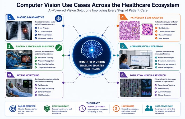
The technical ground under all of this is shifting fast, and the direction matters for anyone making long-term investment decisions in clinical AI.
From Narrow AI to Generalist Models
Traditional medical CV was narrow — one model, one task, painstakingly trained on thousands of manually labeled images. That model is changing. Two developments are driving the transition:
Foundation Models. Meta’s Segment Anything Model (SAM) and its medical adaptation MedSAM — trained on over 1.5 million image-mask pairs — represent the move toward generalist, task-agnostic architectures. MedSAM’s 0.914 Dice score in vertebrae segmentation suggests these broad models will soon rival domain-specific specialists in many clinical tasks. On the architecture side, Vision Transformers (ViTs) like “VerFormer” are already outperforming conventional CNNs on multicenter benchmarks, introducing vertebrae-aware attention mechanisms that improve performance across diverse imaging conditions.
Self-Supervised Learning (SSL). The chronic bottleneck in medical AI has always been labeled data — someone has to manually annotate thousands of images before a supervised model can learn anything. SSL lets models learn meaningful representations from unlabeled data, dramatically reducing that annotation burden. The next evolution — Task-Aligned SSL — fine-tunes this self-supervised learning toward specific clinical goals, combining the scalability of unsupervised training with the precision of task-specific optimization.
Vision-Language Models: Images That Explain Themselves
Perhaps the most consequential frontier right now is the Vision-Language Model (VLM) — multimodal systems that don’t just analyze an image but generate structured clinical language describing what they find. Think of Gemini 2.5 applied to a chest X-ray: not just “abnormality detected in the right lower lobe” but a coherent, contextual description that integrates visual findings with clinical history.
The first foundation-model-powered clinical devices are already receiving FDA clearance, enabling zero-shot learning that adapts to new hospital environments in weeks rather than the years that traditional supervised training required.
Pair VLMs with radiomics — the extraction of quantitative imaging features invisible to the human eye — and the predictive power becomes remarkable. Multimodal fusion models now forecast 3-month functional recovery scores with an AUC of 0.71, predict proximal junctional kyphosis with 72.5% to 100% accuracy, and identify cage subsidence risk with an AUC of 0.81. These are not research curiosities anymore. They are the emerging standard of data-driven perioperative planning.
How to Actually Integrate Computer Vision Into Clinical Workflows
Great technology dies in a silo. Here’s the practical roadmap for making CV work in the real world rather than just in a vendor demo.
Step 1: Start With Site-Specific Prospective Validation
Before you deploy at scale, validate on your patient mix. Confirm performance across your scanner configurations and imaging protocols. Check subgroup performance explicitly — not just overall accuracy — because aggregate metrics can hide serious demographic disparities that only surface in deployment.
Step 2: Solve Interoperability on Day One
The number-one reason CV deployments fail isn’t the algorithm — it’s integration. Diagnostic data lives locked in legacy DICOM and PACS systems. If your AI cannot talk to your existing infrastructure, it becomes a siloed application that adds clicks, adds friction, and accelerates exactly the clinician burnout it was supposed to reduce.
The fix is standards-based connectivity from the start. Build your AI infrastructure to communicate with PACS, RIS, and VNA systems using DICOMweb for modern imaging data access and FHIR for structured health data exchange. Done right, this transforms CV from a pilot project into a genuine operational force multiplier that slots into the workflows clinicians already own.
Step 3: Confront Algorithmic Bias Before It Scales
State-of-the-art models have a documented tendency to underdiagnose pathologies in underserved populations. Trained on narrow, unrepresentative datasets, CV doesn’t just inherit existing healthcare disparities — it formalizes and amplifies them across gender and ethnicity at machine speed. This is not a theoretical concern. It is a documented pattern in published literature, and it is the primary reason site-specific validation is a moral obligation, not just a technical best practice.
Step 4: Make Privacy-First Performance Non-Negotiable
If your deployment involves monitoring or any video-based application, implement privacy protections designed for clinical performance — not just legal compliance. Validate accuracy after privacy transforms like face blurring have been applied. The LookDeep Health data demonstrates this is achievable. Require the same demonstration from any vendor you consider.
Step 5: Govern Updates, Drift, and Retraining
Deploy with a performance monitoring plan in place from day one. Track model accuracy over time against your live patient population. Establish clear retraining and re-approval workflows for when data distributions shift — which they will, as equipment gets upgraded, patient demographics change, and clinical protocols evolve. Document post-market monitoring responsibilities explicitly.
Step 6: Build the Financial Case Honestly
The economics of medical CV deserve straight talk:
- There are still no established CPT codes for most AI-enabled clinical services, which makes reimbursement pathways murky for many applications.
- Deployment demands significant capital expenditure for acquisition, integration, and ongoing maintenance.
- Real-world cost-effectiveness data is sparse. One theoretical model estimated the incremental cost-effectiveness of AI vertebral-fracture screening at $72,085 per quality-adjusted life year (QALY).
The organizations that win in this space will be the ones that build rigorous, honest ROI models before committing — not the ones dazzled by a vendor demo. Clinical economics should be anchored to measurable outcomes: reduced complication rates (like the spine rod fracture example), reduced time-to-treatment, and documented reductions in clinician workload — not projected savings from theoretical efficiency gains.
Frequently Asked Questions
Q1: How many AI-powered medical devices has the FDA authorized?
A: As of the end of 2025, the FDA has authorized 1,451 AI-enabled medical devices, including a record 295 clearances during 2025 alone. Radiology accounts for approximately 76% of all cumulative clearances, reflecting the specialty’s early and sustained adoption of imaging-based AI.
Q2: What is the validation gap in healthcare AI?
A: The validation gap refers to the significant disconnect between regulatory clearance and clinical proof. Roughly 90% of FDA-cleared AI systems rely on retrospective data or have no public clinical evidence, and only 2.4% have been validated through randomized controlled trials. This means a cleared device can still underperform in your specific clinical environment — making site-specific prospective validation essential before deployment.
Q3: Can computer vision accurately monitor patients if their faces are blurred for privacy?
A: Yes. LookDeep Health’s Model v5 achieved an F1-score of 0.91 even with patient faces fully obscured, demonstrating that HIPAA-compliant privacy protections and clinical-grade monitoring performance are simultaneously achievable with the right model architecture.
Q4: How does computer vision improve spine surgery outcomes?
A: CV supports spine surgery across the full perioperative continuum — from preoperative segmentation and alignment measurement to intraoperative navigation and postoperative hardware monitoring. Commercially deployed CV-enabled custom implant planning has reduced rod fracture rates from a historical 9% to 2.2%. Alignment measurement tools like IB Lab SQUIRREL agree with specialist radiologists to within 0.16 degrees.
Q5: How does AI phase recognition improve surgical workflows?
A: CV systems can automatically classify the active stage of a surgical procedure, such as gallbladder retraction during laparoscopic cholecystectomy, enabling real-time safety alerts, automated documentation, and objective skill assessment — without requiring manual annotation of surgical video.
Q6: When do the EU AI Act requirements apply to medical software?
A: The comprehensive high-risk AI obligations for medical devices under the EU AI Act become legally binding on August 2, 2027. Organizations operating in or selling into EU markets should treat this as an active integration deadline, not a future compliance checkbox.
Q7: What is the difference between 510(k) clearance and clinical validation?
A: The 510(k) pathway grants clearance based on substantial equivalence to an existing predicate device — it does not require proof of improved clinical outcomes. Clinical validation, particularly through prospective randomized controlled trials, demonstrates real-world performance on actual patient populations. Only 2.4% of cleared AI devices have cleared that higher bar.
Q8: How do Vision-Language Models support clinical reporting?
A: VLMs like Gemini 2.5 integrate visual feature extraction with natural language generation, enabling systems that don’t just flag abnormalities but describe them in structured clinical language. Applied to radiology and pathology, this supports automated draft reporting and multimodal reasoning across images and clinical records — reducing documentation burden while maintaining physician oversight.
The Road Ahead: Data-Driven Medicine Is Already Here
For all the technical terminology, the story of computer vision in healthcare is really about a fundamental shift in how medicine gets practiced — from experience-based judgment to evidence-backed, computationally augmented precision.
The tools are no longer speculative. They’re triaging urgent scans, guiding surgeons’ hands in real time, watching over ICU patients between rounds, and predicting which patients will develop complications before those complications appear. The FDA’s 1,451 clearances prove the market has arrived. The 2027 EU AI Act deadline proves regulation is catching up.
But arrival isn’t maturity. The gap between a cleared device and a clinically proven tool remains wide, and that gap is where expensive mistakes get made. The winners in this next chapter won’t be the organizations that adopt the most AI. They’ll be the ones that adopt it wisely — demanding prospective evidence over vendor promises, integrating on open standards rather than proprietary silos, confronting bias before it scales, and building financial models grounded in measurable clinical outcomes rather than theoretical efficiency.
Computer vision won’t replace the radiologist, the surgeon, or the nurse. What it will do — when implemented with rigor and honesty — is give every one of them access to the full weight of collective clinical intelligence, processed faster and more consistently than any human team working alone.
That’s not a small thing. It’s a redefinition of what clinical practice is capable of.